Measuring the gut microbiome and its potential to influence wellbeing is now a reality. However, our gut is a large organ and the prevalence of microorganisms and the ability to measure them can vary depending on their location in the gut. So, what exactly can be measured by a stool sample and from where?
When we refer to our gut or gastrointestinal tract, we are referring to the path our food follows during digestion which encompasses the mouth, oesophagus, stomach, small intestine and large intestine (or colon).
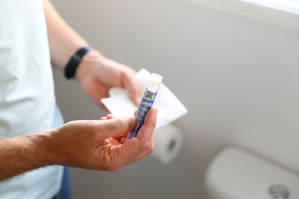
 where the gut microbiome is measured by a Microba metagenomic stool test")
Gut environments and how they relate to the microbiome
Each section of the gut has a very different environment which encourages the growth of different communities of microorganisms. For example, the small intestine is more acidic (due to bile acids) and has higher levels of oxygen and antimicrobial compounds than the large intestine1. It tends to be dominated by fast-growing bacteria that can tolerate these conditions. In contrast, the large intestine has fewer antimicrobial compounds, lacks oxygen, and undigested food (e.g. fibre, excess protein) travels slower1. This encourages the growth of dense populations of bacteria that can process undigested food without oxygen.
The large intestine houses the highest number of bacteria compared to any other part of the gut2.
Where the gut microbiome is found
When people refer to the gut microbiome, generally they mean the microbial community located in the lower part of the large intestine because stool is being used for the measurement. However, stool primarily captures the microbes that are living in the lumen of the large intestine, which is the space where undigested food passes. Studies have shown that a different group of microbes live in the mucus layer lining the large intestine, and a stool sample will not capture this community1.
Want to get to know the community of bacteria living in your large intestine? Find out more here.
Are there parts of the gut microbiome that a stool sample cannot measure?
Although there can be some overlap of bacterial species among different parts of the gut, in general, a stool sample is not representative of the microbiome in the stomach, small intestine, higher up in the large intestine or the mucus layers. A biopsy is required to investigate the microbiome of these areas. Therefore, a stool sample will not suffice when it comes to detecting conditions in other parts of the gut such as small intestinal bacterial overgrowth (SIBO).
The gut microbiome and how it can influence health and wellbeing
The microbiome of stool samples is widely researched because it is much easier to obtain a stool sample than a biopsy sample. This research has found that the stool microbiome of individuals with a variety of physical and mental disorders is different compared to healthy people,3–5 indicating it can be a useful measure of general health.
Although it is unknown yet if differences observed in the gut microbiome are a cause or consequence of the disease, it is a common finding that healthy people have a greater microbial diversity compared to unhealthy people6,7
Additionally, specific bacterial species and bacterially produced metabolites are commonly observed in the stool of unhealthy individuals with specific disease states.
Findings such as these suggest that in the future, we may be able to use the stool microbiome to non-invasively screen for different diseases. It could be that in the next decade when you visit your GP, a microbiome screen will be a normal part of your check-up!
Discover personal health insights through your gut microbiome. Learn more here.
This microbiome test is not intended to be used to diagnose or treat medical conditions. A full disclaimer is available here.
References
1). Donaldson, G. P., Lee, S. M., & Mazmanian, S. K.
Gut biogeography of the bacterial microbiota.
Nature Reviews Microbiology, 14(1), 20 (2016). Doi: 10.1038/nrmicro3552
2). Sender, R., Fuchs, S., & Milo, R.
Revised estimates for the number of human and bacteria cells in the body.
PLoS biology, 14(8), e1002533 (2016). Doi: 10.1371/journal.pbio.1002533
3). Jie, Z., Xia, H., Zhong, S. L., Feng, Q., Li, S., Liang, S., ... & Zhang, D.
The gut microbiome in atherosclerotic cardiovascular disease.
Nature communications, 8(1), 845 (2017). Doi: 10.1038/s41467-017-00900-1
4). Feng, Q., Liang, S., Jia, H., Stadlmayr, A., Tang, L., Lan, Z., ... & Su, L.
Gut microbiome development along the colorectal adenoma–carcinoma sequence.
Nature communications, 6, 6528 (2015). Doi: 10.1038/ncomms7528
5). Marchesi, J. R., Adams, D. H., Fava, F., Hermes, G. D., Hirschfield, G. M., Hold, G., ... & Thomas, L. V.
The gut microbiota and host health: a new clinical frontier.
Gut, 65(2), 330-339 (2016). Doi: 10.1136/gutjnl-2015-309990
6). Valdes, A. M., Walter, J., Segal, E., & Spector, T. D.
Role of the gut microbiota in nutrition and health.
BMJ, 361, k2179 (2018). Doi: 10.1136/bmj.k2179
7). Sommer, F., Anderson, J. M., Bharti, R., Raes, J., & Rosenstiel, P.
The resilience of the intestinal microbiota influences health and disease.
Nature Reviews Microbiology, 15(10), 630 (2017). Doi: 10.1038/nrmicro.2017.58
8). Koh, A., De Vadder, F., Kovatcheva-Datchary, P., & Bäckhed, F.
From dietary fiber to host physiology: short-chain fatty acids as key bacterial metabolites.
Cell, 165(6), 1332-1345 (2016). Doi: 10.1016/j.cell.2016.05.041
9). Lin, H. V., Frassetto, A., Kowalik Jr, E. J., Nawrocki, A. R., Lu, M. M., Kosinski, J. R., ... & Marsh, D. J.
Butyrate and propionate protect against diet-induced obesity and regulate gut hormones via free fatty acid receptor 3-independent mechanisms.
PloS one, 7(4), e35240 (2012). Doi: 10.1371/journal.pone.0035240
10). Bourassa, M. W., Alim, I., Bultman, S. J., & Ratan, R. R.
Butyrate, neuroepigenetics and the gut microbiome: can a high fiber diet improve brain health?.
Neuroscience letters, 625, 56-63 (2016). Doi: 10.1016/j.neulet.2016.02.009